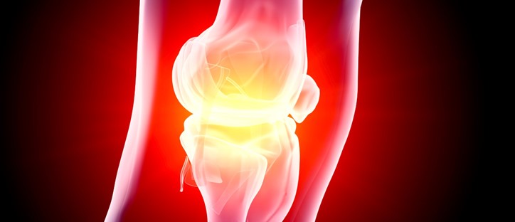
Arthritis is joint inflammation followed by joint destruction. It is the most common cause of disability in America. In 2005, the National Arthritis Data Workgroup revealed that 21% of Americans, over 46 million people, have arthritis or another rheumatic condition. Over 66% of these patients are under 65 years of age. Additionally, in the next 20 years, the number of patients with arthritis is expected to rise by 40% to 67 million people. Arthritis is now epidemic and a significant amount of research, including pharmacologic and device interventions, are aimed at the prevention and mitigation of arthritic pain. Imaging plays a central role inassessing cartilage integrity and the success or failure of these interventions.
Assessment of Arthritis
Arthritis is joint inflammation followed by joint destruction. It is the most common cause of disability in America. In 2005, the National Arthritis Data Workgroup revealed that 21% of Americans, over 46 million people, have arthritis or another rheumatic condition. Over 66% of these patients are under 65 years of age. Additionally, in the next 20 years, the number of patients with arthritis is expected to rise by 40% to 67 million people. Arthritis is now epidemic and a significant amount of research, including pharmacologic and device interventions, are aimed at the prevention and mitigation of arthritic pain. Imaging plays a central role in assessing cartilage integrity and the success or failure of these interventions.
The central feature of arthritis is hyaline cartilage loss. Cartilage is an avascular tissue that covers the surface of articulating bone in synovial joints. It has very little capacity for regeneration. Once fractured, fibrillated, or eroded, cartilage does not engender a robust reparative response. Thus, the preservation of cartilage is of utmost importance in preventing the progression of arthritis. In some cases, areas of chondral damage may be partially repaired or mitigated through such procedures as microfracture, cartilage grafting, and chondrocyte implantation. Once a patient undergoes such a cartilage reparative surgical technique, imaging plays a central role in assessing the success of such a procedure.
Traditionally, cartilage integrity has been inferred by assessment of joint space narrowing on plain film radiography. The cartilage itself is non-calcified and thus invisible on radiographs. However, such imaging, while exquisitely displaying the late changes of cartilage loss such as joint space narrowing, sclerosis, and hypertrophic bone spurring, are woefully inadequate in demonstrating early changes of cartilage loss such as surface fibrillation and changes in cartilage ultrastructure that indicate early cartilage degeneration. It is precisely these early changes that are crucial to recognize in order to implement prompt pharmacologic or surgical intervention.
High Field Magnetic Resonance Imaging
In the last 5 years, advances in magnetic resonance (MR) imaging have improved visualization of hyaline cartilage, allowing much more detailed assessment of both cartilage morphology as well as an indication of cartilage ultrastructural quality.
MR magnets are typically described in terms of magnet strength. Most magnets across the U.S. are rated at 1.5 Tesla. This describes the strength of the magnetic field used to acquire images. In general, the stronger the magnetic field, the better the image quality and image resolution. Approximately 5 years ago, MR manufactures began to produce commercially available 3.0 Tesla (high-field) magnets. These machines have provided a significant leap in radiologists’ ability to image cartilage, both morphologically as well as qualitatively.
The increased magnetic field provided by high field magnets provides an opportunity to significantly increase the spatial resolution afforded by routine MR imaging. Cartilage is typically a thin structure that covers the ends of bones at synovial joints. It may range from less than 1mm in thickness as in the small joints of the fingers and wrist, to 4 to 5mm in thickness as in the knee. Simply stated, increases in spatial resolution afforded by high field MR, when applied appropriately, allow the radiologist to see the cartilage better. Three-dimensional post processing of acquired images also allows better quantification and visualization of cartilage integrity. Surface fibrillation and fissuring, cartilage delamination, and chondral defects are much better visualized and therefore better diagnosed due to these advances in imaging technology.
The holy grail of cartilage imaging is to detect early changes in cartilage quality that indicate early cartilage degeneration before there is progression to frank cartilage loss. Interventions, both surgical and pharmacologic depending upon the underlying arthritis, are aimed at preserving cartilage before there is progression to cartilage loss. Hyaline cartilage has a zonal structure. The thin superficial layer contains densely packed collagen (fibrils) which are oriented parallel to the articular surface. Next is a transitional zone that contains obliquely oriented collagen that gradually changes with depth to become perpendicular. The deepest and largest zone of cartilage abuts the subchondral bone and is termed the radial zone. Because of the different orientation and packing of the cartilage fibrils in the different zones, the water content of the cartilage varies, being slightly higher in the superficial zone than the deeper radial zone. This difference in cartilage water content may be imaged using water sensitive MR pulse sequences. With early cartilage degeneration, before there are morphologic changes in cartilage appearance, there are ultrastructural changes in collagen fibril packing and density. This leads to alterations in water content. High resolution MR imaging, specifically water sensitive T2 sequences, may visually demonstrate such alterations in water content and thereby provide a reflection of cartilage quality within the joint.
The knee is the most common joint to undergo T2 cartilage mapping. This may be applied to either the medial or lateral compartments or the patella. Cartilage maps may be obtained in any plane, but usually are in the axial, sagittal, or coronal planes. Imaging times for each plane acquired are 3 to 5 minutes. The resulting T2 cartilage map is displayed using a color-coded overlay in which good quality, low water content cartilage is red to orange and poorer quality, high water content cartilage is green and blue in color. This map is then compared to high spatial resolution images of the morphologic appearance of the hyaline cartilage to assess for anatomic changes in cartilage integrity. The combination of morphologic assessment and ultrastructural assessment provides a comprehensive picture of the quality of the hyaline cartilage throughout the joint.
Summary
Cartilage preservation is the key endpoint for therapies targeting the management of arthritis. Such therapies include pharmacologic agents for both inflammatory and degenerative arthritides as well as devices and surgical techniques aimed at preserving joint stability. Magnetic resonance imaging, with the advent of high field 3.0 Tesla magnets, now affords the capacity to assess hyaline cartilage changes in their earliest stages before progression to frank cartilage loss and the need for joint replacement. As new interventions are developed to slow the progression of joint inflammation and joint destruction in arthritis, 3.0 Tesla MR imaging with T2 cartilage mapping will play a central role in assessing the ability of these interventions to preserve hyaline cartilage.
Volume 4, Issue 8: Guidance For Sponsors: The Advances of High Field Magnetic Resonance Imaging in the Assessment of Arthritis
Originally written by legacy Intrinsic Imaging Medical Director
Contact WCG Imaging to discuss your trial’s imaging needs
We have the team, therapeutic expertise, technology, and ISO-certified quality management systems to provide imaging core lab services to our clients worldwide. Complete the form to get started.
