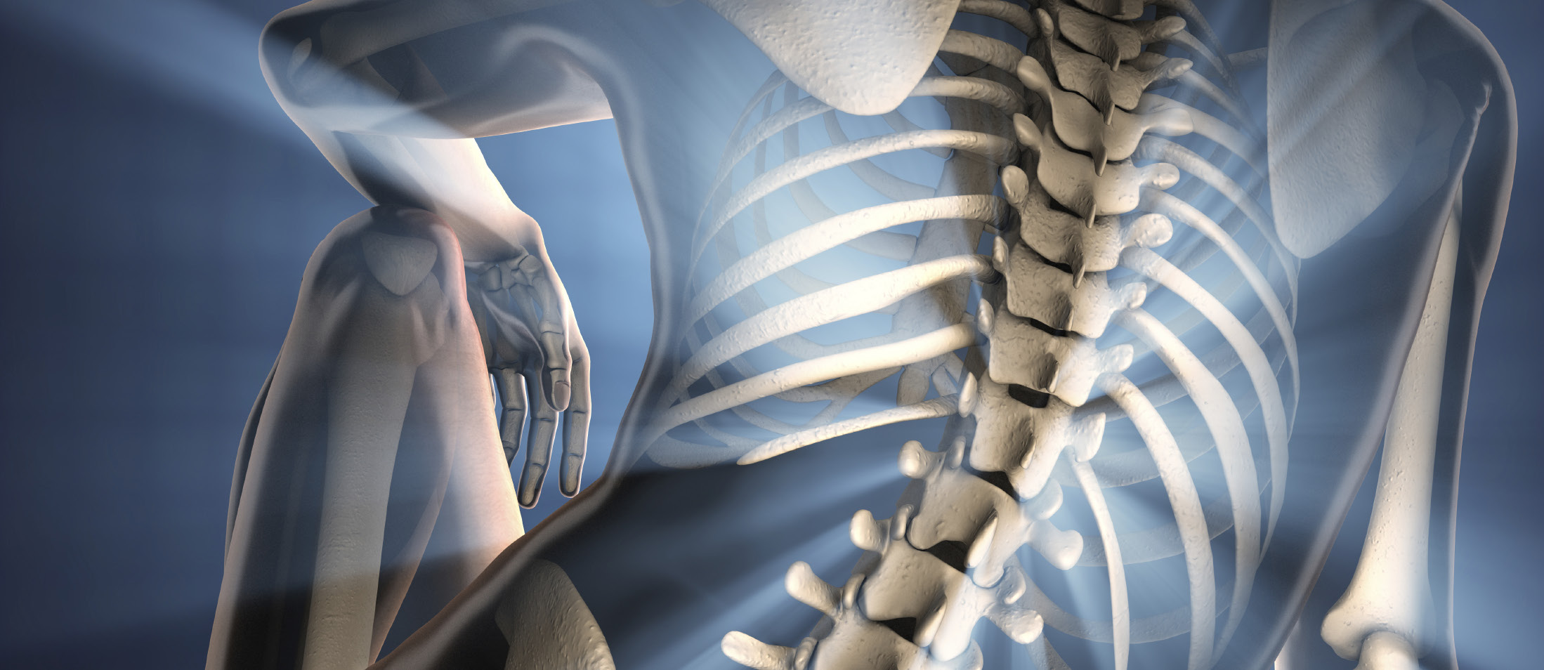
Deformities of the spine, in which the spine is abnormally curved or aligned, can often seriously impact mobility, cause debilitating pain and negatively affect a patients overall quality of life. Patients with complex spinal deformities often require continuous pain management, spinal bracing, or in advanced cases, surgical intervention.
Deformities of the spine, in which the spine is abnormally curved or aligned, can often seriously impact mobility, cause debilitating pain and negatively affect a patients overall quality of life. Patients with complex spinal deformities often require continuous pain management, spinal bracing, or in advanced cases, surgical intervention.
One of the more frequent spinal deformities in children and adults is scoliosis, a lateral curvature of the spine.
In children, scoliosis has many etiologies including idiopathic, congenital, developmental, neuromuscular, and tumor associated. The vast majority of cases of scoliosis in children are idiopathic, further sub-classified as infantile, juvenile, and adolescent types, with an overall incidence in young people of 5 to 7%.
In the adult, there are many causes of spinal deformity, with the most common being idiopathic adolescent scoliosis which has become worse during adulthood due to gradual deterioration of the spine (adult idiopathic scoliosis). A second class of adult spinal deformity, Adult degenerative scoliosis, begins “de novo” in an adult patient without a previous adolescent curve.
Medical Imaging
Scoliosis and spinal deformity imaging is targeted at determining both the anatomy of the underlying curvature as well as attempting to assess its progression. Radiography, computed tomography (CT), and magnetic resonance imaging (MR) all play a role in scoliosis assessment. In the adolescent, MR is important for assessing secondary etiologies for scoliosis such as developmentally anomalous vertebral bodies or underlying osseous or neurologic pathology. In the adult, MR provides a tremendous amount of information regarding degree of disc pathology, facet arthritis, spinal canal stenosis, and neural foraminal compromise. This is crucial information for therapeutic intervention, be it minimally invasive or surgical.
CT examination also plays an important role in scoliosis evaluation. CT depicts the three-dimensional anatomy of the scoliotic curvature both in adults and adolescents. It is unsurpassed in its ability to demonstrate the true degree of lateral bending as well as rotational changes in the axial plane. Additionally, CT is able to provide detailed information regarding post-surgical complications such as hardware malplacement or failure. This includes such complications as rod or pedicle screw fracture, pedicle screw malpositioning, bone fracture, failed fusion, and pseudarthrosis formation. However, CT is limited in its ability to provide dynamic information as well as its ability to provide cost-effective longitudinal analysis for assessing disease progression.
Radiography remains the primary imaging modality in the assessment of scoliosis and other complex spinal deformities. It provides an overall assessment of the spinal axis in the coronal and sagittal planes. Right and left lateral bending views allow dynamic assessment and the ability to distinguish a structural curve from non-structural curves. Radiography is limited by its two-dimensional planar technique. Thus, the rotatory component of scoliosis is comparatively poorly imaged and poorly assessed. However, as radiography has the historical imaging advantage, nearly all of the quantitative criteria used to assess scoliosis and its progression are radiographically based. Current clinical trials targeted to scoliosis and other spinal deformity imaging continue to rely upon the radiograph as the gold standard for imaging assessment.
The quantitative criteria used to assess a spinal curvature were developed for assessment of idiopathic adolescent scoliosis, but are applicable to all forms of spinal deformity. The Cobb angle is the most commonly used quantitative assessment of the scoliotic curve, both clinically and in clinical trials. It is defined as the angle formed by the intersection of two lines, one parallel to the endplate of the superior vertebral body with maximal tilt, and the other parallel to the endplate of the inferior vertebral body with maximal tilt. These are defined as the end vertebra. The apex of curvature is defined as the most lateral point of curvature and may be a vertebral body level or disc level. Neutral vertebrae are defined as vertebrae outside the span of the curve that show no evidence for rotation (symmetric pedicles) on frontal radiographs. Measurement of the Cobb angle is limited in that it is performed on a two-dimensional radiograph of a three dimensional deformity. The more severe the curvature, the more prominent is its rotational component. This places the plane of maximal lateral curvature further away from the imaged frontal coronal plane and may lead to an underestimation of the true Cobb angle. Longitudinal analysis of changes in Cobb angle over time, used to assess progression of curvature, require consistent positioning from one time point to the next. Additionally, Cobb angle assessment should be performed at the same levels from one examination to the next. Variations in Cobb angle assessment due to acquisition technique can range from 2° to 7°. The interobserver variation can reach approximately 5° to 10°. Given these ranges of error, curve progression is measured in increments of 5° and a progressive curve is one in which there is an increase of 5° between successive examinations.
Frontal bending views allow the differentiation of major (structural) curves from minor (nonstructural) curves. This differentiation is important for surgical planning. A major curve is usually defined as one with a Cobb angle that remains greater than 25° on ipsilateral frontal bending views. The central sacral vertebral line (CSVL), drawn through the center of the sacrum and perpendicular to a tangential line drawn across the tops of the iliac crests, and the plumb line, drawn vertically from the center of the C7 vertebral body, are used to assess coronal balance on frontal views and sagittal balance on lateral views respectively. Stable vertebrae are defined as the farthest cranial vertebra bisected by the CSVL.
Standardization in Clinical Trials
The standardization of measurements in clinical trials for spinal deformities and spine devices is of paramount importance. The utilization of classification systems aids in standardizing the parameters used by radiologists in blinded settings. There is not a widely accepted classification for adult scoliosis or adult deformity. In idiopathic adolescent scoliosis, the Lenke classification is the most commonly used classification system. It has three components: curve type, lumbar modifier, and thoracic sagittal modifier. Curve type is divided into three regions based upon the apex level: T1-T3 (proximal thoracic), T3- T12 (main thoracic), and T12-L4 (thoracolumbar). All three regions are assessed as to the presence of major or minor curves. Curves may be major (structural), minor and structural, or minor and nonstructural (some minor curves may be structural). This leads to six discrete curve types in the Lenke system based upon the location of the major curve and minor curves: Main thoracic, double thoracic, double major, triple major, thoracolumbar/lumbar, and thoracolumbar/lumbar-main thoracic. A lumbar spine modifier (A, B, or C) is then assigned based upon the position of the lumbar curve apex vertebra in relation to the CSVL. If the CVSL lies between the pedicles, it is termed “A”, if the CSVL touches the pedicle it is scored a “B”, and lateral to pedicle is assigned a “C”. The last step is to assign a sagittal modifier based upon the degree of kyphosis between T5 and T12. When the angle of kyphosis is less than 10°, the modifier “-“ is assigned; when the angle is 10-40°, the modifier “N” is assigned; and when the angle is greater than 40°, the modifier “+” is assigned. The angle of kyphosis is measured using the Cobb method. The complete Lenke classification therefore requires standing frontal and lateral radiographs as well as frontal leftward and rightward bending views.
Imaging plays a central role in the evaluation of scoliosis and other complex spinal deformities, both in initial evaluation, assessment of disease progression, as well as the success of surgical and nonsurgical interventions. CT and MR provide different types of anatomic detail with MR important in initial evaluation to assess for causative factors or complications from the spinal deformity, and CT useful in the demonstration of the true three-dimensional appearance of the complex spinal curve as well as potential complications related to surgery. MR is also very informative in adult scoliosis, demonstrating the degree of disc degeneration and arthritis that underlies the progressive deformity. Despite the anatomic detail afforded from these cross sectional imaging techniques, radiography remains the mainstay for longitudinal scoliosis assessment in both the clinical and research arenas. Radiography, despite its limitations in assessing a three-dimensional disease with a two-dimensional modality, nevertheless provides a reproducible, consistent, and validated means to assess spinal curvature and curve progression.
Volume 6, Issue 4: Guidance for Sponsors: Complex Spinal Deformities: Medical Imaging in Clinical Trials
Originally written by legacy Intrinsic Imaging Medical Director
Contact WCG Imaging to discuss your trial’s imaging needs
We have the team, therapeutic expertise, technology, and ISO-certified quality management systems to provide imaging core lab services to our clients worldwide. Complete the form to get started.
