The aging of the baby boomer generation is postulated to inspire a host of refinements and novel devices geared toward orthopedic interventions. These patients demand a more active lifestyle and rigorously pursue medical therapies toward that end. Orthopedic clinical research encompasses a diversity of initiatives ranging from improvements in joint reconstruction hardware, spinal fusion and spinal motion preserving interventions, to novel biologics ranging from articular and fibrocartilage repair to tendon and ligament regrowth and reconstruction. Regulatory and market acceptance of these novel therapies requires rigorous quantitative and qualitative assessment to determine efficacy, safety, and longevity of these orthopedic advancements.
Orthopedic Device Assessments
The aging of the baby boomer generation is postulated to inspire a host of refinements and novel devices geared toward orthopedic interventions. These patients demand a more active lifestyle and rigorously pursue medical therapies toward that end. Orthopedic clinical research encompasses a diversity of initiatives ranging from improvements in joint reconstruction hardware, spinal fusion and spinal motion preserving interventions, to novel biologics ranging from articular and fibrocartilage repair to tendon and ligament regrowth and reconstruction. Regulatory and market acceptance of these novel therapies requires rigorous quantitative and qualitative assessment to determine efficacy, safety, and longevity of these orthopedic advancements.
Imaging endpoints are crucial in the assessment of orthopedic devices and surgical therapies in both the clinical and research fields. Imaging provides an abundance of qualitative and quantitative data useful in the evidence based assessment of experimental orthopedic interventions. Orthopedic investigational devices often are quite novel. An intimate understanding of all aspects of the device including engineering, material composition, surgical placement technique, and device positioning is important in the design of imaging protocols tailored to assessing the success or failure of such device. Musculoskeletal radiologists with significant experience in longitudinal device assessment are best suited to protocol design and subsequent data interpretation as they most commonly possess the conceptual expertise with orthopedic hardware assessment that can be translated to devices which have frequently never been imaged before.
Dynamic and Static Evaluations
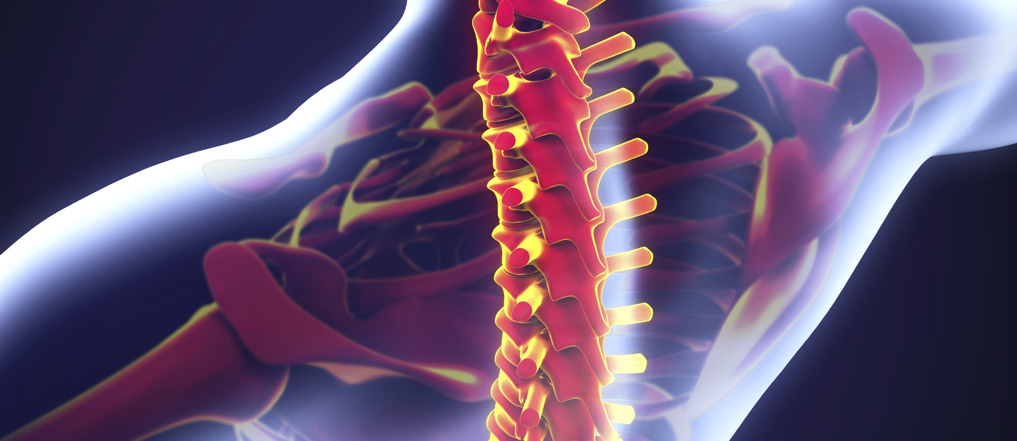
The imaging assessment of orthopedic devices and biologics may be broadly divided into dynamic and static evaluations. By definition, orthopedic devices are subject to motion and the stresses therein. Dynamic assessment is most commonly performed in the setting of spinal devices using radiography or dynamic fluoroscopy. Flexion and extension radiographs or dynamic fluoroscopic imaging of spinal motion produces an imaging data set that may be processed using specific software to generate a large amount of quantitative data reflecting spinal motion and alignment changes. Such data includes disc height changes, angular motion assessment, and evaluation of changes in spondylolisthesis. Software analysis has been shown to provide reproducible data accurate to less than 1 degree and 1 mm. Additionally, the presence of metallic hardware does not routinely preclude the identification of the anatomic landmarks used in quantitative motion assessment. Cross-sectional imaging modalities such as magnetic resonance imaging and computed tomography are not typically used in dynamic motion analysis as patients are typically confined to one position during image acquisition.
The static assessment of orthopedic devices is accomplished using an array of imaging modalities including radiography, computed tomography (CT), and magnetic resonance imaging (MRI). Each modality provides unique information reflecting the in vivo changes in bone and soft tissues as well as the device itself. In the case of metal hardware, radiography provides a broad assessment of the investigational device and potential device related complications. Such complications include device migration, subsidence, device disassembly, and bone changes such as fracture, remodeling, and hypertrophic bone formation or ankylosis.
CT performs a similar role as radiography, but imparts much increased sensitivity and specificity to detection of all the above complications, often revealing findings not detectable on plain film radiography due to superimposition of anatomy. CT also allows for accurate measurements of both device and adjacent bone anatomy that can be performed in multiple planes. Additionally, CT provides improved, although suboptimal, assessment of soft tissues adjacent to orthopedic devices. CT protocols must be optimized to the investigational device, especially with respect to those devices containing metallic components. In general, maximizing kilovoltage potential (kVp) and milliampere- seconds (mAs) is employed to minimize the beam hardening artifact and subsequent streaking that degrade CT image quality. Additionally, protocols using thin slice technique with overlap of slice acquisition provide a robust data set that can be constructed into images in which the bone-device interface is well demonstrated. Finally, use of multi-detector scanners allows a larger amount of tissue to be imaged and therefore a larger degree of slice acquisition overlap that also increases image quality in the setting of metallic hardware.
The assessment of soft tissue changes and complications adjacent to orthopedic hardware is best performed using MRI techniques. MRI allows the detection of soft tissue changes related to device placement including synovitis, periarticular fluid collections, particle disease, arthrofibrosis, muscle injury, bone stress changes, and changes in cartilage morphology and quality. MRI is also the optimal imaging modality for assessing novel biologics involved in articular cartilage and soft tissue reconstruction methods, providing detailed information related to biologic graft and cartilage graft positioning, integrity, incorporation and adjacent bone changes. In the spine, MRI detects bone marrow changes adjacent to device placement as well as soft tissue changes specific to the spine such as disc degeneration, disc herniation, epidural hematoma, infection, and CSF leaks.
Historically, MRI has been limited in its use in the evaluation of metal containing devices or arthroplasty hardware due to susceptibility artifacts and distortion of the regional magnetic field. These artifacts rendered images useless. However, recent advancements in MRI techniques have minimized these effects and opened up the use of MRI in the evaluation of metallic hardware in both the clinical and research settings.TheMARS(metalartifactreductionsequence)protocol adds a compensation gradient to correct the inhomogeneity induced intensity changes around the device. SEMAC (slice encoding for metal artifact reduction) eliminates metal- induced distortions by encoding each excited slice against metal-field inhomogeneities to suppress both in-plane distortions as well as distorted excitation profiles associated with through plane distortions. Both are quite effective in allowing visualization and evaluation of anatomy in close proximity to metallic hardware and are of rapidly increasing use in clinical practice.
Summary
As novel orthopedic devices enter clinical evaluation, imaging modality, acquisition and analysis must be tailored to the needs of the trial. Each modality provides different,but complementary qualitative and quantitative data. Advancements in CT and MRI techniques afford these modalities a greater role in precise evaluation of orthopedic devices. Additionally, the use of radiography is enhanced by the application of software evaluation techniques, especially in the case of spinal motion analysis.
Volume 4, Issue 5: Guidance For Sponsors: Orthopedic Device Assessments: Medical Imaging Challenges in Clinical Trials
Originally written by legacy Intrinsic Imaging Medical Director
Contact WCG Imaging to discuss your trial’s imaging needs
We have the team, therapeutic expertise, technology, and ISO-certified quality management systems to provide imaging core lab services to our clients worldwide. Complete the form to get started.