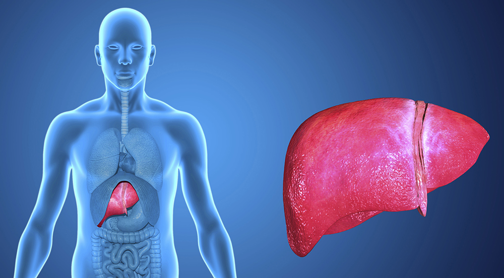
Magnetic resonance elastography is an emerging MRI technology that provides sensitive and semi-quantitative assessment of tissue stiffness. The most promising clinical application for MR Elastography is the assessment of liver stiffness as a surrogate marker of liver disease and fibrosis. Liver fibrosis is the primary pathologic process in cirrhosis which affects millions of Americans and causes significant morbidity and mortality. The incidence of liver disease and cirrhosis is increasing, especially cirrhosis related to chronic viral hepatitis and nonalcoholic fatty liver disease (NAFLD). When incorporated into a comprehensive liver MRI exam, elastography provides a sensitive assessment for early diffuse liver disease and a semi- quantitative assessment for stage and progression of fibrosis.
Magnetic Resonance Elastography
Magnetic resonance elastography is an emerging MRI technology that provides sensitive and semi-quantitative assessment of tissue stiffness. The most promising clinical application for MR Elastography is the assessment of liver stiffness as a surrogate marker of liver disease and fibrosis. Liver fibrosis is the primary pathologic process in cirrhosis which affects millions of Americans and causes significant morbidity and mortality. The incidence of liver disease and cirrhosis is increasing, especially cirrhosis related to chronic viral hepatitis and nonalcoholic fatty liver disease (NAFLD). When incorporated into a comprehensive liver MRI exam, elastography provides a sensitive assessment for early diffuse liver disease and a semi-quantitative assessment for stage and progression of fibrosis.
Management of patients with liver disease such as chronic viral hepatitis or NAFLD involves careful monitoring for the development and progression of liver inflammation, fibrosis, and cirrhosis. This is usually achieved through a combination of physical exam, laboratory testing, and imaging. Imaging may include abdominal ultrasound, CT, or MRI exams. However, all of these tests may remain normal during the early stages of liver disease. The liver does not demonstrate gross morphological abnormalities such as nodular contour or segmental atrophy until advanced stages of fibrosis; complications of cirrhosis such as portal venous hypertension, splenomegaly, and ascites also indicate advanced disease. Therefore, in patients with potential early disease, liver biopsy is often used to assess for presence of active liver inflammation and fibrosis. Liver biopsy is an expensive and invasive procedure with associated risks. Also, liver biopsy suffers from sampling error, sometimes underestimating or overestimating the actual degree of liver disease.
MR Elastography has been developed by Dr. Richard Ehman and colleagues at the Mayo Clinic as a noninvasive mechanism of evaluating tissue stiffness. Dr. Ehman’s group and others have demonstrated that patients with liver disease have progressively increased stiffness of their liver tissue which can be detected and quantified with MR Elastography, years before the development of gross anatomic abnormalities of liver size and shape that are detectable by other imaging modalities.
MR Elastography requires specialized equipment and software, which are included with some new MRI scanners and can be added to many installed scanners. The necessary equipment is relatively simple: a small disc-shaped transducer is placed over the patient’s upper abdomen by the technologist at the start of the exam, and the transducer is connected through a flexible plastic tube to a speaker-like device, which creates low frequency sound waves at a fixed frequency of approximately 40-90 Hertz. These sound waves pass through the transducer and cause small vibrations within the patient’s abdomen, without discomfort to the patient. These vibrations create pressure waves within the liver which are detectable through sensitive MR sequences. Specifically, MR Elastography employs phase contrast MR sequences which are sensitive to the direction and amplitude of tissue motion. In patients with chronic liver disease, as the liver tissue becomes stiffer, the wavelength of the pressure waves becomes longer. This can be visualized in color-coded images, and specialized image analysis software can measure the wavelengths and provide quantitative estimates of tissue stiffness, through- out the liver or within a specific region of interest. Tissue elasticity results are reported in kilopascals (kPa). The entire MR Elastography acquisition can be performed in 1 minute, with negligible increase in overall length of the MRI exam.
Improved Detection of Early Liver Fibrosis
Studies have demonstrated that MR Elastography provides highly reliable measurements, with little test-retest variation. Normal subjects demonstrate consistent liver stiffness measurements with mean of 2.2 kPa and standard deviation of 0.3 kPa, corresponding to normal soft liver tissue. Patients with increasing degrees of liver fibrosis demonstrate a direct correlation between elastography measurements and stage of fibrosis defined in liver biopsy specimens. Importantly, patients with early liver disease
and stage 0 or stage 1 fibrosis demonstrate elevated liver stiffness measurements of approximately 3.5-4.0 kPa. Using a cut-off value of 2.93 kPa, Yin et al. achieved remarkable sensitivity of 98% and a specificity of more than 99% for differentiating any stage of liver fibrosis from normal liver tissue (reference below). Using a cut-off value of 4.89 kPa, these researchers were able to differentiate early disease with stages 0-1 fibrosis from more advanced disease with stages 2-4 fibrosis, with 86% sensitivity and 85% specificity. These results represent a marked improvement over other available tests for early liver disease and will enable greatly improved detection of early liver fibrosis in patients at risk.
MR elastography will allow clinicians to more sensitively and accurately identify the presence and progression of hepatic fibrosis in patients with chronic liver disease, especially the large number of patients with viral hepatitis (5 million Americans) and nonalcoholic fatty liver disease (31 million Americans). MR elastography results will potentially help guide clinicians’ decisions about when to perform invasive liver biopsy and how and when to adjust patient therapy. As improved therapies become available, including therapies targeted at slowing, preventing, or even reversing hepatic fibrosis, the highly repeatable and quantitative measurements obtained by MR Elastography will provide accurate assessment of treatment responses.
Summary
Some therapies are already in use for treatment of chronic viral hepatitis, such as Interferon and Ribavirin, and clinicians may begin to use elastography results to assess the necessity and efficacy of these treatments. Within the larger number of patients with NAFLD, approximately 10- 20% will develop active liver inflammation and fibrosis, termed nonalcoholic steatohepatitis (NASH), with significant risk of progression to cirrhosis. Currently there are no specific therapies for NASH, but there is tremendous research interest in this direction. Experimental treatments for NASH include trials of newer antidiabetic medications that increase insulin sensitivity and may reduce liver injury. NIH-sponsored studies of these medications in patients with NASH are underway, including trials of rosiglitazone, pioglitazone, and metformin. There are many other pharmaceutical agents in development for the prevention and treatment of liver fibrosis. MR elastography will play an important role in future trials of these agents, providing noninvasive and quantitative measurements of liver fibrosis, potentially complementing or partially replacing liver biopsy in assessment of patients’ stage and progression of liver disease.
References
Yin et al., Assessment of hepatic fibrosis with magnetic resonance elastography, Clin Gastroenterol Hepatol. 2007 Oct; 5(10):1207-13.
Volume 4, Issue 1: Guidance For Sponsors: Magnetic Resonance Elastography: A Novel Technique for Evaluation of Liver Disease
Originally written by legacy Intrinsic Imaging Medical Director
Contact WCG Imaging to discuss your trial’s imaging needs
We have the team, therapeutic expertise, technology, and ISO-certified quality management systems to provide imaging core lab services to our clients worldwide. Complete the form to get started.
