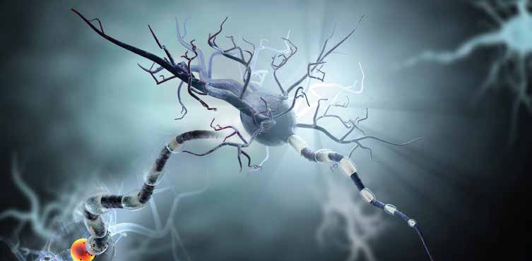
Neurodegenerative Disorders
The diagnosis of movement disorders and neurodegenerative diseases can be problematic for clinicians and radiologists alike. There is considerable overlap in the early presentation of these diseases especially when tremor is absent or atypical in character. Routine anatomic imaging is insensitive and early imaging findings are typically absent or non-specific.
After Alzheimer Disease (AD), Parkinson Disease (PD) is the second most common neurodegenerative disorder, affecting approximately 1 million Americans and 4 million worldwide. PD affects 0.5-1.0% of individuals between 65 and 69 years old and 1-3% older than 80.
The cause of idiopathic PD is the degeneration of neurons which connect the subtantia nigra in the midbrain to the corpus striatum of the basal ganglia, the so called nigrostriatal pathway. These neurons control the release of the neurotransmitter dopamine to receptor cells in the basal ganglia which affect motor function. Pre-synaptic dopamine transporter proteins are located on the cell membranes of these terminal neurons which continuously reuptake dopamine from synaptic clefts after the completion of interaction with dopamine receptors on the post-synaptic neuron. Non-degenerative Parkinsonian syndromes may be drug induced or vascular as a result of ischemic insults to the pathway.
Imaging of the Dopamine Pathway
Functional nuclear medicine imaging of the dopamine pathway using DaTscan and Fluorodopa offers renewed hope in categorizing these diseases as degenerative Parkinsonian syndromes or non-degenerative in etiology.
DaTscan is a trademark radiopharmaceutical used for striatal dopamine transporter imaging with single-photon emission computed tomography (SPECT) technique. More specifically, the DaTscan agent is the I-123 isotope of iodine bound to fluoropropyl-carboxymethyl-iodophenyl nortropane (FP-CIT). It binds reversibly to human recombinant dopamine transporters and is blocked by dopamine reuptake inhibitors. The dopamine transporter is thus a surrogate marker for dopaminergic nigrostriatal neurons.
Fluorodopa, also known as F-DOPA, is a fluorinated form of L-DOPA. It is synthesized as a fluorine-18 analog for use as a radiotracer in positron emission tomography (PET) imaging.
Dopamine in circulation does not cross the blood-brain barrier of the central nervous system, however, L-DOPA is carried across by the neutral amino acid transport system. L-DOPA is then converted to dopamine in the brain by L-aromatic amino acid decarboxylase. Dopamine is stored in pre-synaptic intraneuronal vesicles and released when the nigrostriatal nerve cell fires.
Both DaTscan SPECT and F-DOPA PET imaging assess the pre-synaptic dopaminergic system at functionally similar points on the dopamine pathway. DaTscan images correlate with dopamine transporter density in the corpus striatum while F-DOPA images correlate with the activity of the decarboxylating enzyme and the storage capacity of dopamine in vesicles located in these same structures. The end product of both studies is images of the corpus striatum and interpretation of the relative intensity of radio-isotope uptake.
PET imaging requires less than one hour of incubation time between isotope injection and imaging. The spatial resolution of PET is moderately superior to that of SPECT. The lack of practical commercial availability of the PET isotope has limited its use to academic centers with on-site or near site cyclotrons. Furthermore, the F-18 isotope has a short half-life and its binding to L-DOPA is a rigorous process.
In contrast, the I-123 isotope for DaTscan SPECT imaging has a relatively long half-life and is more readily available. Imaging of the I-123 DaTscan agent requires incubation of 3 to 4 hours.
Parkinson Disease vs Atypical Parkinsonian Syndromes
Early in PD, dopamine transport is down regulated to preserve pre-synaptic dopamine. This enhances DaTscan SPECT imaging sensitivity. Conversely, the decarboxylating enzyme can be upregulated as a compensatory mechanism to produce more pre-synaptic dopamine. Thus, F-DOPA PET imaging may be falsely negative early in PD. Both tests are highly effective in differentiating PD and the atypical Parkinsonian syndromes (APS) of Multiple System Atrophy, Progressive Supranuclear Palsy, Corticobasal Degeneration and Dementia with Lewy Bodies from non-degenerative Essential Tremor (ET) and drug-induced Parkinsonism.
It is generally accepted that the motor symptoms of PD occur after 60-80% of dopamine producing neurons are lost and that loss proceeds at a rate of 6-13% per annum. Degeneration occurs in dopaminergic cells which originate in the substantia nigra and terminate in the corpus striatum. The disease process produces loss of striatal dopamine and the dopamine transporters that collect spent dopamine from the synaptic cleft. The loss of neurons in the striatal terminals proceeds from lateral to medial, thereby affecting the posterior putamen before the caudate head. PD and APS typically produce asymmetric motor symptoms. The imaging abnormalities are correspondingly variable and can be unilateral, bilateral or bilaterally asymmetric. In ET and drug-induced Parkinsonism, there is no nigrostriatal dopaminergic cell loss, therefore, imaging by either modality is normal.
DaTscan SPECT imaging provides an in vivo marker of dopaminergic neuronal degeneration which helps differentiate PD and APS from ET, drug-induced Parkinsonism and other non-Parkinsonian syndromes. There is no clinical biomarker for the pathologic substrate of PD, the intraneuronal inclusion of Lewy bodies in the substantia nigra compacta. Clinical history, physical examination and a positive response to L-dopamine remain the standards for diagnosis. PD is generally characterized by an asymmetric rest tremor, slow movements (bradykinesia), stiffness (cogwheel rigidity) and problems with walking or balance (postural instability). A positive clinical diagnosis is established by bradykinesia and one additional cardinal feature plus a positive response to dopamine therapy. In ET, tremor is the primary symptom, typically symmetrical and produced with action or upon standing.
Sometimes PD patients have no tremor or a postural tremor that mimics ET. Occasionally ET patients have an asymmetric tremor, rest tremor or cogwheeling rigidity that mimics PD. The overtreatment of non-PD patients with anti-Parkinsonian medications is estimated between 15-47%. Post-mortem confirmation of a non-PD diagnosis in a hospital study of patients treated for PD has been reported between 10-24%.
DaTscan SPECT imaging can be a valuable tool in reducing the overdiagnosis of PD. A negative SPECT study in the presence of tremor has a negative predictive value of 87.5%. Conversely, an abnormal DaTscan has a positive predictive value for PD or APS of virtually 100%.
Conclusion
As the prevalence of movement disorders increases in our aging population at great personal and financial costs to society, disease documentation becomes more important than ever. New and improved medical imaging places radiology practices at the forefront of diagnosis. Functional imaging of the dopamine pathway will continue to play an increasing role in clinical diagnosis, appropriately qualifying patients for clinical trials and in documenting disease prior to invasive device implantation or targeted ablation. It is essential for cutting edge radiology practices and core trial labs to become familiar with the newest functional molecular imaging tools at our disposal.
Furthermore, since both Parkinson Disease and atypical Parkinsonian syndromes can produce cognitive decline, functional imaging modalities such as DaTscan SPECT and F-DOPA PET will become increasingly beneficial for the accurate diagnosis of these patients in conjunction with volumetric MRI, FDG-PET and amyloid PET imaging of the brain.
References
- Broski S, Hunt C, Johnson G, Lowe V, Peller P. Structural and Functional Imaging in Parkinsonian Syndromes. Radiographics 2014; 34: 1273-1292.
- Marshall V, Reininger C, Marquardt M, Patterson J, Hadley D, Oertel W, Benamer H, Kemp P, Burn D, Tolosa E, Kulisevsky J, Cunha L, Costa D, Booij J, Tatsch K, Chaudhuri K, Ulm G, Pogarell O, Hoffken H, Gerstner A, Grosset D. Parkinson’s Disease is Overdiagnosed Clinically at Baseline in Diagnostically Uncertain Cases: A 3-Year European Multicenter Study with Repeat [123I]FP-CIT SPECT. Movement Disorders 2009; 24(4): 500-508.
- Booij J, de Bruin K, van Royen E, Speelman J, Horstink M, Sips H, Dierckx A, Versijpt J, Decoo D, Van Der Linden C, Hadley D. Doder M, Lees A, Costa D, Gacinovic S, Oertel W, Pogarell O, Hoeffken H, Joseph K, Tatsch K, Schwarz J, Ries V. Accurate Differentiation of Parkinsonism and Essential Tremor Using Visual Assessment of [I123]-FP-CIT SPECT Imaging: The [I123]-FP-CIT Study Group. Movement Disorders 2000; 15(3): 503-510.
- Nanni et al. 18F-DOPA PET and PET/CT. Journal of Nuclear Medicine 2007; 48(10): 1577-1579. Hauser R, Grosset D. [I123]FP-CIT (DaTscan) SPECT Brain Imaging in Patients with Suspected Parkinsonian Syndromes. Journal of Neuroimaging 2012; 22(3): 225-230.
Volume 5, Issue 3: Guidance For Sponsors: Functional Imaging of the Dopamine Pathway
Originally written by legacy Intrinsic Imaging Medical Director
Contact WCG Imaging to discuss your trial’s imaging needs
We have the team, therapeutic expertise, technology, and ISO-certified quality management systems to provide imaging core lab services to our clients worldwide. Complete the form to get started.
