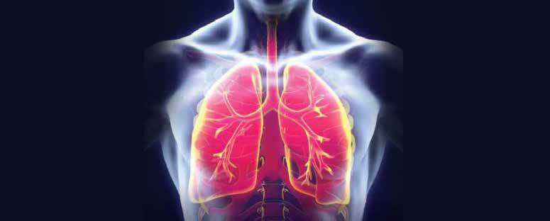
Lung Cancer
Lung cancer is the leading cause of cancer death across the globe, claiming more lives than the next three most common cancers combined (colon, breast and pancreatic). In 2012, the estimate for new cases of lung cancer reached 226,160, with 164,770 deaths accounting for 27% of all cancer deaths. The five-year survival rate is low at 16%, often due to most patients having advanced disease at the time of presentation.
Non-small cell lung cancer (NSCLC) accounts for some 85% of lung cancer diagnoses. Fortunately for this subset of patients, surgery can often be curative, but only if presentation is within stages 1 or 2. However, it is a sad fact that 70% of presentations are at stages 3 or 4, and so much attention has recently been given to screening – and specifically the use of computed tomography (CT) imaging – in an attempt to detect a larger number of these cases at an early point in the disease process. For patients with late-stage disease, the oncologic community has turned to novel therapies.
Diagnoses and Therapy
Advances in our understanding of lung cancer have led to better diagnostic and therapeutic techniques. The last decade has seen an appreciation of the genetic basis of cancer, with research identifying highly effective targeted therapies for particular genomic changes. State-of-the-art management now involves personalised treatment using genomic cancer information.
Given the various lung cancer subtypes, treatment can be chosen based on different mutation status, and a genomic analysis can be used in tailoring ongoing therapy. Doctors can utilise molecule-specific agents that work against an aberrant molecular pathway to both maximise therapeutic efficacy and limit the toxicity from the treatment. Within the lung cancer genomic arena, the most therapeutically important mutations to guide treatment have been:
Epidermal Growth Factor Receptor (EGFR)
In NSCLC, the discovery of the mutations of the EGFR tyrosine
kinase domain is associated with a signifi cant
response to the EGFR-TKI inhibitors. The three most commonly used targeted agents are gefi tinib (Iressa), erlotinib (Tarceva) and cetuximab (Erbitux). These are now part of mainstay therapy, having been granted
approval for the treatment of NSCLC in many countries based on recent Phase 3 randomised controlled trials and
the recommendation of the American Society of Clinical Oncology.
EML4-ALK Gene Fusion
Relevant in adenocarcinoma and in patients who have never smoked, the EML4-ALK fusion is present in 3-6% of cases. For the treatment of lung cancer that demonstrates presence of the EML4-ALK fusion, crizotinib (Xalkori) has emerged as an effective therapy.
Ki-ras2 Oncogene (KRAS)
The KRAS mutation is most common in patients who smoke or have done in the past. Several agents in development or in clinical trials to target genes like KRAS will soon come to market.
Tumour Assessment
Diagnostic devices, and methods for detection and staging, have evolved towards less invasive and more accurate tools. Endobronchial ultrasound and electromagnetic navigation have been the main focus of invasive techniques and the evolution of CT for non-invasive imaging.
Imaging assessment to response of tumour therapy possesses validated techniques and is fundamental in shaping cancer treatments. The effectiveness and accuracy of tumour response assessment has a direct impact here. A deep understanding of modalities is crucial in taking forward imaging biomarkers. Within this discipline, considerable effort has been devoted to establishing standardised measurement techniques.
The conventional assessment on routine imaging such as CT has typically revolved around single planar measurements by radiologists, without significant automation. The WHO criteria and the Response Evaluation Criteria in Solid Tumours (RECIST 1.0 and 1.1) are often used, but novel measurement parameters are emerging as imaging techniques advance. Several of these include fl uorodeoxyglucose positron emission tomography (FDG-PET), novel tracer PET, magnetic resonance imaging (MRI), and CT tumour volume and perfusion, as well as diffusion-weighted MRI. These techniques are already fi nding application in various therapeutic areas and trials – and now also in lung cancer.
Limits of Criteria
As the most widely used methodologies, the details of
implementing WHO and RECIST criteria are well-described in the literature. The problem with these criteria, however, mainly concerns the reliance on size measurements alone – ignoring the biology of the tumours with heterogeneous growth patterns, notably in NSCLC and other genomically modulated tumours treated with targeted therapies.
Firstly, RECIST criteria may not show expected conventional progression in genetically modulated tumours as typically seen in other tumours. The biology of certain subsets of NSCLC can show a continued growth in size, but can still possess tumour tissue that is sensitive to novel therapies and would continue to respond to treatment. There are now several trials in patients with NSCLC that harbour EGFR mutations, which are beginning to demonstrate delayed responses when treated beyond what is considered progression by RECIST criteria. The growth pattern of NSCLC can show a rapid initial response and then slow progression over many months – a pattern not well-suited to RECIST-based measurements.
Secondly, tumours do not grow in a predictable spherical pattern and often the incumbent methodologies can either over- or under-represent changes in tumour growth. In addition, these methodologies do not encompass the physiology of tumours where, in certain cases, there can be stability in size but show central necrosis or cavitation, representing response.
There has also been increasing scrutiny of the validity of current measurement methodologies. Investigators have demonstrated that there is statistically signifi cant variability in interpretation utilising both the WHO and RECIST criteria when interpreting CT scans on NSCLC patients, both in single dimension and bi-dimensional measurements (1,2). In those sets of studies, RECIST 1.1 was found to provide superior performance than RECIST 1.0, which shows better performance than WHO criteria. With all these emerging questions, the imaging community is beginning to look at alternate methodologies and modalities.
PET Imaging
PET is a functional imaging technique that produces a multiplanar image of functional processes in the body, and has long featured in oncologic and, specifically, lung imaging. While imaging primarily relies on CT, integrated PET/CT has been found to be more effective than PET alone, CT alone, or a combination of correlating PET and CT.
The primary agent in PET imaging is FDG which, as an analogue of glucose, allows an understanding of metabolism in tumours. Within the lung, FDG-PET has been instrumental in differentiating benign and malignant nodules, as well as the staging of cancer. Initial work has been conducted in NSCLC to evaluate correlation between the EGFR mutation and FDG avidity on PET.
Furthermore, PET imaging in oncology applications is starting to utilise alternative radioactive tracers that can be tagged to various molecules. Radiolabelled ligands are being developed to study molecular-targeted therapy and the effects of various anti-cancer agents on end organs. There is also the possibility of labelling cancer therapeutics themselves.
In NSCLC, studies have been performed to label gefitinib and erlotinib, which have been shown to accumulate in models of lung tumour and lymph nodes – identifying lesions that were not visible using conventional FDG for PET imaging.
Volumetric Measurements
Given the heterogeneity in tumour growth in three dimensions, considerable work has been expended on tools that enable the measurement of tumour volume as a biomarker for evaluation. With the advent of multi-detector CT scanners, imaging has evolved to isotropic voxel acquisition, where accurate volumes can be measured given the quality of image acquisition. Many studies have shown that utilising this in lung tumours is more reproducible and standardised than using a uni-dimensional or bi-dimensional approach (1,3,4). This leads to reduced variance between readers and over time.
Cutting-edge work in tumour volume has shown that volumetric tumour measurements during genetic-based therapies for NSCLC could be used to distinguish tumours with the mutation from those without the mutation (5). Given the continued development of techniques, this looks set to play a crucial role in assessing tumour response in the future. However, it is important to note that, currently, tumour volume is not a validated biomarker, and the technique needs further investigation.
CT and MRI Perfusion
Advances in CT and MRI acquisition technology have enabled the use of multiple images obtained through a lesion during a rapid time sequence in the administration of intravenous contrast. This methodology, termed ‘perfusion imaging’, permits the evaluation of uptake or the contrast material over time within a lesion. The technique uses, at a cellular level, the concepts of permeability and blood volume and, if standardised, could provide information about tumour biology. Due to the technical nature of this evaluation, additional software is required at present, and there has been little agreement between software packages to establish perfusion as a valid biomarker.
With regards to use of this technique in evaluating the response of tumours to therapy in the lung, preliminary work has shown that baseline blood flow and blood volume in patients with partial response by RECIST criteria were higher, compared to patients with stable or progressive disease (6).
Perfusion-based criteria has been taking shape, notably in hepatocellular carcinoma (HCC), for several years. The HCC community has understood that anatomic tumour response measurements could be misleading in relation to molecular-targeted therapies or locoregional therapies in HCC. Thus, in 2008, a group of experts from the American Association for the Study of Liver Diseases established a set of guidelines that incorporated the concept of viable tumour tissue, showing uptake in arterial phase of contrast-enhanced imaging techniques, and formally amended RECIST. In future, similar amendments may be required for lungspecific therapies.
IDiffusion-Weighted MRI
Diffusion MRI is a method established in the mid-1980s, primarily for the detection of cerebral stroke, and is now being used in various parts of the body, including the liver and lung. It allows the mapping of the diffusion process of molecules – mainly water – non-invasively within the body.
The water molecule diffusion pattern reveals microscopic details about tissue characteristics. Within the lung imaging sphere, diffusion-weighted MRI has recently been investigat- ed as a possible biomarker for tumour evaluation. Initial work has suggested that particular changes in diffusion parame- ters could potentially classify patients that may have longer progression-free survival and overall survival. Ultimately, this technique may provide an in-depth understanding of tumour biology, as it has in other processes within the body, but it could be diffi cult to use in the actual evaluation of treatment response.
New Window
The development of novel genetic-based therapies for NSCLC has opened a new window to previously late-stage disease. The radiology community has taken the challenge of establishing new imaging biomarkers to assist in assessing the response to these therapies with existing tools, as well as creating novel tools and techniques to improve understanding of tumour biology at a cellular level. However, from measurement techniques to new modalities, imaging CROs will require high level expertise to perform trials evalu- ating these therapies. Implementing incorrect biomarkers or imaging strategies can lead to wrong assessments of failure for therapies, and result in loss of valuable treatment options.
References
- Zhao B et al, Evaluating variability in tumor measurements from same-day repeat CT scans of patients with non-small cell lung cancer, Radiology 252(1): pp263-272, 2009
- Nishino M et al, New Response Evaluation Criteria in Solid Tumors guidelines for advanced non-small cell lung cancer: Comparison with original RECIST and impact on assessment of tumor response to targeted therapy, Am J Roentgenol 195(3): W221-228, 2010
- Choi H et al, CT evaluation of the response of gastrointestinal stromal tumors after imatinib mesylate treatment: a quantitative analysis correlated with FDG PET fi ndings, Am J Roentgenol 183(6): pp1,619-1,628, 2004
- Zhao B et al, Lung cancer: Computerized quantifi cation of tumor response-initial results, Radiology 241(3): pp892-898, 2006
- Zhao B et al, A pilot study of volume measurement as a method of tumor response evaluation to aid biomarker development, Clin Cancer Res 16(18): pp4,647-4,653, 2010
- Wang J, Wu N, Cham MD and Song Y, Tumor response in patients with advanced non-small cell lung cancer: Perfusion CT evaluation of chemotherapy and radiation therapy, Am J Roentgenol 193(4): pp1,090-1,096, 2009
Volume 5, Issue 2: Guidance For Sponsors: Advances in Imaging of Non-Small Cell Lung Cancer (NSCLC)
Originally written by legacy Intrinsic Imaging Medical Director
Contact WCG Imaging to discuss your trial’s imaging needs
We have the team, therapeutic expertise, technology, and ISO-certified quality management systems to provide imaging core lab services to our clients worldwide. Complete the form to get started.
